Nuclear Instruments and Methods in Physics Research B 268, 3243 (2010)
Swift heavy ion irradiation-induced microstructure modification
of two delta-phase oxides: Sc4Zr3O12
and Lu4Zr3O12
M. Tang a,*, P. Kluth b, J. Zhang a,
M.K. Patel c, B.P. Uberuaga a,
C.J. Olson Reichhardt d, K.E. Sickafus a
aMaterials Science & Technology Division, Mail-Stop G755,
Los Alamos National Laboratory, Los Alamos, NM 87545, USA
bDepartment of Electronic Materials Engineering, The Australian
National University, Canberra, ACT 0200, Australia
cHigh Pressure Physics Division, Bhabha Atomic Research
Centre, Mumbai 400 085, India
dTheoretical Division, Mail-Stop B268, Los Alamos National
Laboratory, Los Alamos, NM 87545, USA
ARTICLE INFO
Article history:
Received 2 October 2009
Received in revised form 28 May 2010
Available online 2 June 2010
Keywords:
Cermaic oxides
Irradiation damage effects
Phase transformation
TEM
XRD
Abstract
Swift gold ions (185 MeV) were used to systematically investigate the radiation
damage response of delta
phase compounds Sc4Zr3O12
and Lu4Zr3O12 in the
electronic energy loss regime. Ion irradiation-induced
microstructural modifications were examined using X-ray diffraction (XRD) and
transmission electron
microscopy (TEM). XRD investigations indicate a phase transformation from
ordered rhombohedral to
disordered fluorite (O-D) in both compounds, with the Sc compound transforming
at a higher ion fluence
compared with the Lu compound. This result is consistent with our previous study
on Sc4Zr3O12 and
Lu4Zr3O12 under
displacive radiation environment in which the nuclear energy loss is dominant. High
resolution TEM revealed that individual ion tracks maintain crystalline structure,
while the core region
experiences an O-D phase transformation.
TEM observations also suggest that for the doses in which
the tracks overlap, the O-D phase transformation occurs across the entire ion range.
1. Introduction
2. Experimental procedure
3. Results and discussion
4. Summary
References
1. Introduction
The radiation damage behavior of various complex oxides with
structures related to fluorite (with potential application as host
materials for nuclear waste or as advanced nuclear fuel forms for
the transmutation of minor actinides) has been the focus of many
recent studies
[1,
2,
3,
4,
5].
The fluorite structure (CaF2 type, Fm3̅m),
consisting of a face-centered cubic array of cations with anions in all of
the tetrahedral interstices is adopted by MO2 dioxides (M represents
a metal cation, while O refers to an oxygen anion). Certain
oxygen-deficient fluorite structural derivatives (MO2 - x ), such as
pyrochlore (M4O7)
compounds (more specifically A3+ 2B4+ 2 O7 );
bixbyite (M2O3) compounds (like rare earth sesquioxides); and
δ-phase (M7O12)
compounds (more specifically A3+ 4B4+ 3 O12 ),
exhibit amorphization resistance upon irradiation to very high radiation
dose (pyrochlores [6],
bixbyite [7], and δ-phase
[5,
8]).
The purpose of the study presented here is to investigate radiation
damage effects in Sc4Zr3O12
and Lu4Zr3O12 in the electronic
energy loss regime. Swift heavy ions (SHI), namely, 185 MeV Au
ions were used to simulate radiation damage effects similar to
those induced by fission products in nuclear fuel. Both
Sc4Zr3O12
and Lu4Zr3O12
compounds are known to crystallize in the δ-phase
structure, which is described as a rhombohedral distortion (space
group R3̅
[9, 10])
of the fluorite structure with anionic vacancies
on one of the cubic 3-fold axes. We showed previously
[5,
8] that
these compounds are very resistant to irradiation-induced amorphization,
even at cryogenic temperature (100 K). However, these
δ-phase compounds do undergo an order-to-disorder (O-D) phase
transformation from an ordered δ-phase to a disordered cubic fluorite
structure, in a displacive irradiation environment.
2. Experimental procedure
High purity ZrO2, Lu2O3
and Sc2O3 powders from Alfa Aesar
(99.99% purity) were used to produce sintered polycrystalline
Sc4Zr3O12 and
Lu4Zr3O12 pellets.
Pellet fabrication was performed
using conventional ceramic processing procedures. X-ray diffraction
measurements showed the pristine oxide ceramics to be phase
pure and possess rhombohedral symmetry. The pellets were polished
with alumina lapping films to obtain a mirror finish. In preparation
for ion irradiation, all of these samples were final-polished
using 40 nm colloidal silica slurry (Syton HT50, DuPont AirProducts
NanoMaterials L.L.C, Tempe, AZ) to remove the surface damage
created by mechanical polish.
Irradiations were performed at room temperature with 185 MeV
Au13+ ions over a range of fluencies ranging from 1 × 10¹¹
ions/cm2
to 1 × 10¹³ ions/cm2,
at the Heavy-Ion Accelerator Facility of The
Australian National University, Canberra, Australia. The ion beam
was scanned over an aperture of 3 × 6 mm2, upstream of the
sample, to ensure homogeneity. The beam current was maintained between
5 nA and 10 nA resulting in power densities below
1 W/cm2. The Monte Carlo program
SRIM [11] was used to estimate
electronic and nuclear stopping in SHI irradiated
Sc4Zr3O12 and
Lu4Zr3O12 .
A threshold displacement energy of 40 eV was used for
all target elements (this is an arbitrary assumption). Fig. 1 shows
the results of a SRIM simulation for Sc4Zr3O12 .
The electronic stopping power, (dE/dx)electronic,
exceeds the nuclear stopping power,
(dE/dx)nuclear,
except at the very end-of-range of the ion. Upon entering the solid,
the electronic stopping powers is ~30 keV/nm/ion for
Sc4Zr3O12 and Lu4Zr3O12 (not shown here).
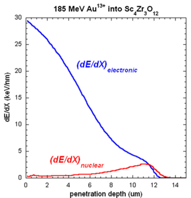 Figure 1:
Monte Carlo simulation estimates of electronic versus nuclear energy loss
for 185 MeVAu ions as function of penetration depth for Sc4Zr3O12.
Figure 1:
Monte Carlo simulation estimates of electronic versus nuclear energy loss
for 185 MeVAu ions as function of penetration depth for Sc4Zr3O12.
|
Irradiated samples were analyzed using both X-ray diffraction
(XRD) and transmission electron microscopy (TEM). XRD measurements
were made using a Bruker AXS D8 Advanced X-ray diffractometer,
Cu-Kα radiation, a graphite monochromator, and θ-2θ
geometry. The diffractometer was equipped with a Göebel mirror
to achieve parallel beam diffraction optics. Irradiated samples were
also prepared in cross-sectional and plan-view geometries for TEM
examination. The SHI irradiation-induced microstructural evolution
was examined using both a Philips CM-30 and a FEI Tecnai
F30 electron microscope, each operating at 300 kV. Microdiffraction
(μD) was used in this study to obtain diffraction patterns from
small sample areas (the electron probe size at the sample was focused
to a diameter of 10-20 nm).
3. Results and discussion
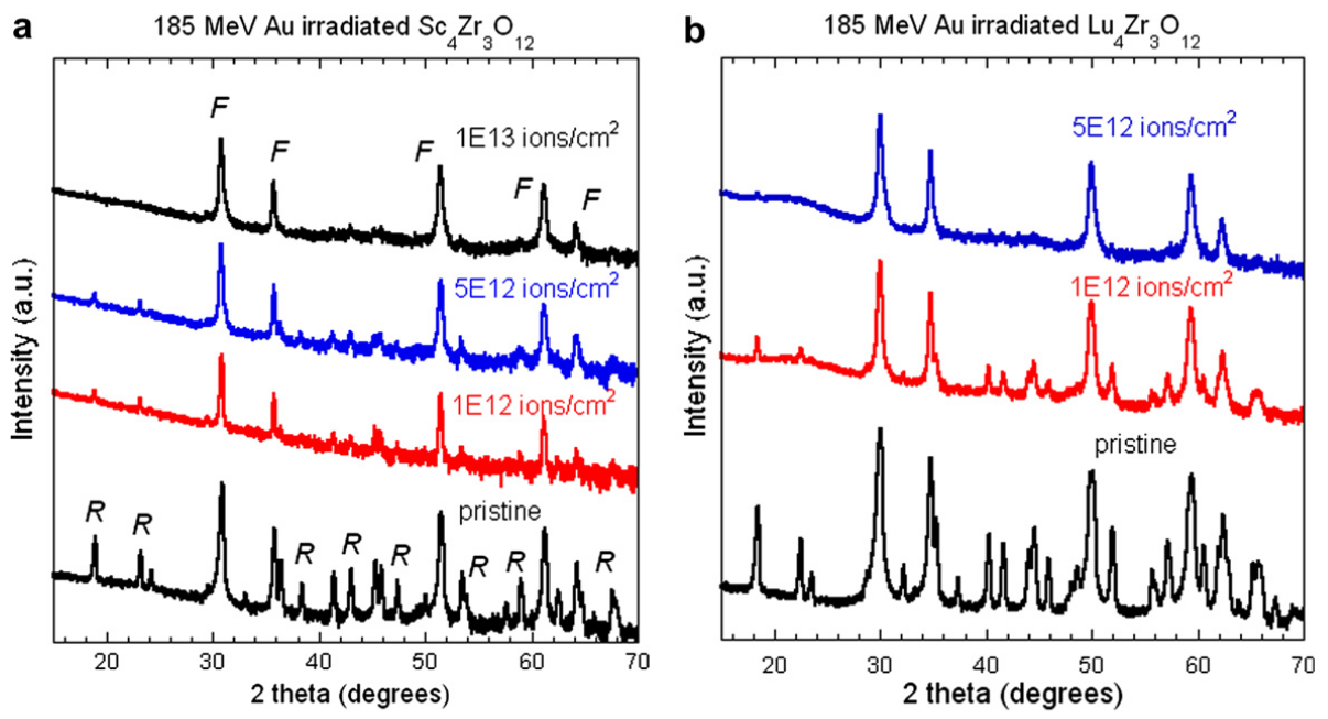
 Figure 2:
X-ray diffraction (XRD) patterns obtained from (a) Sc4Zr3O12
and (b) Lu4Zr3O12
before and after irradiation with 185 MeV Au ions. XRD indicated that both compounds
experience an irradiation-induced order-to-disorder (O-D)
phase transformation, from an ordered rhombohedral to a disordered fluorite.
Plots are shown as logarithmic in
intensity.
Figure 2:
X-ray diffraction (XRD) patterns obtained from (a) Sc4Zr3O12
and (b) Lu4Zr3O12
before and after irradiation with 185 MeV Au ions. XRD indicated that both compounds
experience an irradiation-induced order-to-disorder (O-D)
phase transformation, from an ordered rhombohedral to a disordered fluorite.
Plots are shown as logarithmic in
intensity.
|
Fig. 2a and b are GIXRD patterns obtained from Sc4Zr3O12 and
Lu4Zr3O12
samples before and after 185 MeV Au ion irradiation,
respectively. The initial structure of Sc4Zr3O12
can be described
as an ordered rhombohedral δ-phase. The peaks labeled 'R' in
Fig. 2 represent this rhombohedral, δ-phase. Upon ion irradiation
to the highest fluence of 1 × 10¹³ Au/cm2,
one important observation is that there are no apparent broad diffraction features,
attributable to an amorphous structure. This observation suggests that
no SHI irradiation-induced amorphization occurs in either
Sc4Zr3O12 or
Lu4Zr3O12.
Interestingly, with increasing ion irradiation fluence the weakest
δ-phase R peaks decrease in intensity
more than the four most prominent diffraction peaks (at ~31°,
36°, 52°, 61° 2θ). These R peaks are almost completely absent at
the highest fluence, 1 × 10¹³ Au/cm2 .
The four major diffraction
maxima peaks are associated with the "parent" fluorite structure
(diffraction peaks labeled 'F') in Fig. 2, while the weaker R peaks
are due to the special structural arrangement associated with the
fluorite derivative, δ-phase structure. The absence of the weaker
δ-phase (R) reflections with increasing ion irradiation dose suggests
that Sc4Zr3O12
and Lu4Zr3O12 gradually undergo an O-D
transformation, from an ordered δ-phase structure to a disordered
fluorite structure. One difference between
Sc4Zr3O12
and Lu4Zr3O12
structural evolution with irradiation dose, is that for
Lu4Zr3O12 , all
R peaks associated with δ-phase disappear leaving only F peaks by
a fluence of 5 × 10¹² Au/cm2.
This effect occurs at a lower ion
fluence for Lu4Zr3O12
compared to Sc4Zr3O12 . To summarize, XRD
investigations indicate that SHI irradiation induces an O-D phase
transformation from an ordered rhombohedral (R) to a disordered
fluorite (F) phase in both Sc4Zr3O12
and Lu4Zr3O12 , with the Sc compound
transforming at a higher ion fluence compared to the Lu
compound. This result is consistent with our previous study in
which we compared the radiation damage response of Sc4Zr3O12
and Lu4Zr3O12
under displacive radiation damage conditions (i.e.,
conditions wherein the nuclear energy loss dominates)
[5].
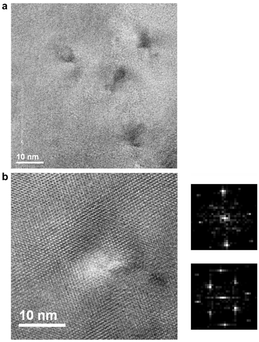
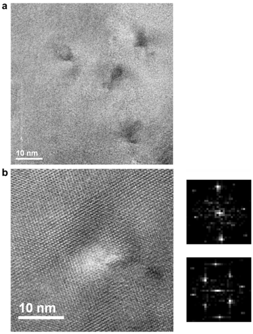 Figure 3:
Plan-view high resolution TEM images obtained from
Sc4Zr3O12
irradiated to a fluence of 1 × 10¹¹ Au/cm2 .
(a) Individual ion tracks are observed with round shapes of
~3-4 nm in diameter; (b) higher magnification than (a), showing an individual ion track
consisting of disordered fluorite core (~3-4 nm). Also shown in (b) are
diffractograms obtained by Fast Fourier Transforms (FFT)
from both the core and matrix.
Figure 3:
Plan-view high resolution TEM images obtained from
Sc4Zr3O12
irradiated to a fluence of 1 × 10¹¹ Au/cm2 .
(a) Individual ion tracks are observed with round shapes of
~3-4 nm in diameter; (b) higher magnification than (a), showing an individual ion track
consisting of disordered fluorite core (~3-4 nm). Also shown in (b) are
diffractograms obtained by Fast Fourier Transforms (FFT)
from both the core and matrix.
|
Fig. 3 shows plan-view, high resolution TEM images obtained
from Sc4Zr3O12
irradiated to a fluence of 1 × 10¹¹ Au/cm2 . These
images were obtained from a sample thinned to reveal the structure
at the irradiated sample surface (corresponding to an electronic
stopping power of ~30 keV/ion/nm). In Fig. 3, single ion
tracks are visible, each with round shapes of ~3-4 nm in diameter
(Fig. 3a). Lattice fringes are clearly resolved inside each ion track.
This indicates that δ-phase Sc4Zr3O12
is not amorphized by the
individual track formation process. Also note in Fig. 3 the strong
dark contrast apparent in some regions. This is most likely caused
by strain contrast due to volume changes within the different
phases. Lattice fringes in Fig. 3b reveal that the cores of ion tracks
in Sc4Zr3O12
possess a different structure compared to the matrix.
Fast Fourier Transform (FFT) analysis suggests that the ions producing
these tracks induced an O-D phase transformation. We
based this conclusion on the partial disappearance of the superlattice
reflections associated with the rhombohedral, δ-phase. High
resolution TEM observations reveal that ion tracks in Sc4Zr3O12
consist of a disordered fluorite structured core surrounded by a
δ-phase matrix.
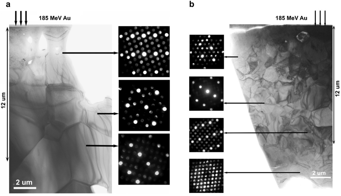
 Figure 4:
Cross-sectional TEM bright field (BF) images and microdiffraction patterns
obtained from (a) Sc4Zr3O12
irradiated to a fluence of 1 × 10¹³ Au/cm2 ;
(b) Lu4Zr3O12
irradiated to a fluence of 5 × 10¹² Au/cm2 .
The decrease in intensity of specific diffraction spots suggests that an
O-D phase transformation occurs within the ion range.
Figure 4:
Cross-sectional TEM bright field (BF) images and microdiffraction patterns
obtained from (a) Sc4Zr3O12
irradiated to a fluence of 1 × 10¹³ Au/cm2 ;
(b) Lu4Zr3O12
irradiated to a fluence of 5 × 10¹² Au/cm2 .
The decrease in intensity of specific diffraction spots suggests that an
O-D phase transformation occurs within the ion range.
|
Fig. 4a and b show cross-sectional TEM images and microdiffraction
(μD) patterns obtained from Sc4Zr3O12
irradiated to a fluence of 1 × 10¹³ Au/cm2
and Lu4Zr3O12 irradiated to a fluence of
5 × 10¹² Au/cm2 , respectively.
At these high ion fluences, it is possible to assess microstructural
change induced by overlapping ion
tracks. In Fig. 4, the μD patterns were obtained from different
depths within the irradiated regions, progressing from the surface
to the ion end-of-ranges, respectively. The $mu;D patterns obtained
from the ion range (~12 um) in Fig. 4a and b are consistent with
a two-phase structure, where both phases are oriented with an epitaxial
relationship with respect to the pristine δ-phase substrate.
The strong reflections in μD patterns are consistent with a cubic
fluorite structure. The weaker reflections are equivalent to those
in the pristine δ-phase substrate pattern (referred to as superlattice
reflections, characteristics of pristine delta phase). The diminishing
intensities of the superlattice reflections associated with the rhombohedral
δ-phase, compared to the stronger, fundamental fluorite
reflections, suggests that the irradiated Sc4Zr3O12
and Lu4Zr3O12
experience O-D phase transformations following Au ion irradiation.
The only μD pattern obtained out of the ion range in Fig. 4b
is clearly consistent with a single phase, the rhombohedral
δ-phase. The TEM observations in Fig. 4 corroborate the XRD
results presented in Fig. 2 indicating that Sc4Zr3O12
and Lu4Zr3O12
remain crystalline, but experience a structural transformation after
overlapping of ion tracks, from an ordered δ-phase to a structure
indistinguishable from a cubic fluorite. Observations by μD revealed
that this O-D phase transformation occurred across the entire
ion range (corresponding to electronic stopping power ranging
from ~30 keV/ion/nm to <5 keV/ion/nm).
The O-D phase transformation in Sc4Zr3O12
and Lu4Zr3O12 has
been observed previously under displacive irradiation damage
conditions, using both heavy ions (300 keV Kr)
[5, 8] and light ions
(200 keV Ne) [12].
Nuclear energy loss dominates the Kr irradiation process,
whereas electronic energy loss plays a greater role
in the total stopping power for Ne. As the disordering process under
displacive conditions, the Sc δ-phase compound transforms at
higher ion fluence compared to the Lu compound. In this study
using swift heavy ions, we observed the same O-D phase transformation
as in the previous displacive damage irradiations, and
once again, we found differences between the irradiation damage
response of Sc4Zr3O12 and
Lu4Zr3O12 . The O-D phase transformation
in δ-phase rhombohedral compounds is normally associated
with high-temperature polymorphic transformation, as revealed
in Temperature-Composition (T-C) phase diagrams. Irradiation-induced
phase transformations to higher temperature polymorphs
have been observed previously in other oxides
[13,
14,
15].
By analogy, the different irradiation response in these two materials
examined in this study (Sc and Lu) may be anticipated by
phase-stability characteristics, as revealed in T-C diagrams. These
T-C phase diagrams
[16] show that a thermally-induced O-D
phase transformation occurs at ~1800 °C in Sc4Zr3O12 and at
~1700 °C in Lu4Zr3O12 .
Our experimental results on irradiation
dose dependence of the O-D phase transformation observed in
Sc4Zr3O12 and
Lu4Zr3O12 appear
to be consistent with the temperature dependence of the
O-D phase transformation observed in
the phase diagram for these two materials.
Based on our XRD measurements, we find that the molecular
density increases in both Sc4Zr3O12
and Lu4Zr3O12 (or the volume
decrease) 0.37% and 0.57%, respectively, upon ion irradiation to
the highest fluence (1 × 10¹³ Au/cm2 )
used in these experiments.
However, the experimental data presented in Lopato et al.
[9] indicates
that there is virtually no change in volume during the O-D
phase transformation in Sc4Zr3O12 .
This is consistent with our
SHI irradiation-induced O-D phase transformation. The 0.37% density
(or volume change) found in our experiments is not statistically
significant. Essentially, we find no change in density or
volume upon transformation.
4. Summary
We performed SHI irradiation experiments under room
temperature on polycrystalline δ-phase compounds,
Sc4Zr3O12
and Lu4Zr3O12 ,
using 185 MeV Au13+ ions. Between a fluence of
5 × 10¹² Au/cm2 and
1 × 10¹³ Au/cm2 , a crystal structure transformation
occurs from an ordered rhombohedral to a disordered fluorite
structure in both compounds, where the Sc compound
transforms at an higher ion fluence compared with the Lu compound.
This transformation seems to be identical to a thermally-induced
O-D transformation that occurs at high temperature in
these compounds. HRTEM observation reveals that disordered
fluorite regions are formed at the core of the ion tracks. The diameters
of these disordered regions in Sc4Zr3O12
is ~3-4 nm diameter. Electron diffraction patterns also suggest that the O-D phase
transformation occurs across the entire ion range in both the Sc
and Lu compounds (corresponding to electronic stopping power
ranging from ~30 keV/ion/nm to <5 keV/ion/nm). No irradiation-induced
amorphization was observed in either Sc4Zr3O12 or
Lu4Zr3O12 .
Acknowledgment
This work was sponsored by the Laboratory Directed Research &
Development-Exploratory Research (LDRD-ER) at Los Alamos National
Laboratory. We thank the staff at the ANU Heavy-Ion Accelerator
Facility for technical assistance. P.K further acknowledges
the support of the Australian Research Council.
References
- [1]
-
W.J. Weber, R.C. Ewing, C.R.A. Catlow, T. Diaz de la Rubia, L.W. Hobbs, C. Kinoshita,
Hj. Matzke, A.T. Motta, M. Nastasi, E.K.H. Salje, E.R. Vance, S.J. Zinkle,
J. Mater. Res. 13 (1998) 1434.
- [2]
-
K.E. Sickafus, L. Minervini, R.W. Grimes, J.A. Valdez, M. Ishimaru, F. Li,
K.J. McClellan, T. Hartmann, Science 289 (2000) 48.
- [3]
-
C. Degueldre, M. Pouchon, M. Dobeli, K. Sickafus, H. Hojou,
G. Ledergerber, S. Abolhassani-Dadras, J. Nucl. Mater. 289 (2001) 115.
- [4]
-
R.C. Ewing, W.J. Weber, J. Lian, J. Appl. Phys. 95 (2004) 5949.
- [5]
-
K.E. Sickafus, R.W. Grimes, J.A. Valdez, A.R. Cleave, M. Tang, M. Ishimaru,
S.M. Corish, C.R. Stanek, B.P. Uberuaga, Nature Mater. 6 (2007) 217.
- [6]
-
J. Lian, X.T. Zu, K.V.G. Kutty, J. Chen., L.M. Wang, R.C. Ewing, Phys. Rev. B
66 (2002) 71.
- [7]
-
M. Tang, P. Lu, J.A. Valdez, K.E. Sickafus, J. Appl. Phys. 99 (2006) 063514.
- [8]
-
J.A. Valdez, M. Tang, K.E. Sickafus, Nucl. Instr. and Meth. B 250 (2006) 148.
- [9]
-
L.M. Lopato, V.P. Red'ko, G.I. Gerasimyuk, A.V. Shevchenko, Neorg. Mater. 27
(8) (1991) 1718.
- [10]
-
H.J. Rossell, J. Solis State Chem. 19 (1976) 103.
- [11]
-
J.F. Ziegler, J.P. Biersack, U. Littmark, The Stopping and Range of Ions in Solids,
Pergamon, New York, 1985.
- [12]
-
J. Zhang, Y.Q. Wang, M. Tang, J. Won, J.A. Valdez, K.E. Sickafus,
J. Mater. Res. 25 (2010) 248.
- [13]
-
S. Hemon, V. Chailley, E. Dooryhee, C. Dufur, F. Gourbilleau, F. Levesque,
E. Paumier, Nucl. Instr. and Meth. B 122 (1997) 563.
- [14]
-
J.A. Valdez, M. Tang, Z. Chi, M.I. Peeters, K.E. Sickafus, Nucl. Instr. and Meth. B
218 (2004) 71.
- [15]
-
M. Tang, P. Lu, J.A. Valdez, K.E. Sickafus, Nucl. Instr. and Meth. B 250 (2006) 142.
- [16]
-
H.M. Ondik, H.F. McMurdie (Eds.), Phase Diagrams for Zirconium and Zirconia
Systems, The American Ceramic Society, Westerville, 1998.
File translated from
TEX
by
TTHgold,
version 4.00.
Back to Home